Dental Health
3D Intraoral Scanner Benefits in Madrid
A 3D intraoral scan creates a detailed digital model of your teeth without traditional impression materials. Here is when it helps—and when other tests are still needed.
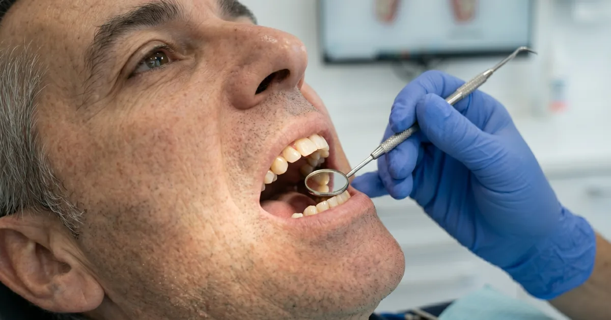
The Debod team’s perspective on 3D intraoral scanners
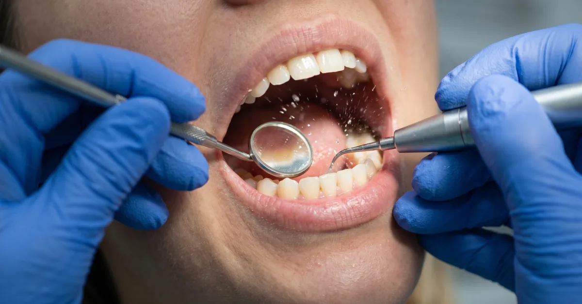
At Debod Dental Clinic in Argüelles, Madrid, a 3D intraoral scanner is part of how we make diagnosis and treatment planning clearer for patients. It is a compact optical camera that records the visible surfaces of teeth, gums and the way the bite meets, then builds a three-dimensional digital model on screen.
For many people, that is a welcome change from impression trays filled with material. No strong taste, less sensation of bulk in the mouth, and no waiting for material to set. But honest dentistry means being precise about what the technology does. A scan is a detailed record, not a diagnosis or a treatment by itself.
It does not remove decay, stop gum disease, correct bruxism or replace a missing tooth. It helps the dentist see, plan and communicate. Think of it like a very accurate map: useful for choosing the route, but it does not drive the car.
Is it normal to wonder whether it involves radiation? No radiation is used by the scanner. Where we need information about roots, bone or hidden decay, we may still recommend an X-ray or a 3D CBCT scan. The first diagnostic visit is the right moment to decide which records are genuinely needed for your case.
The image appears as we work. You can see areas that are difficult to judge in a mirror, and we can explain why a crown, retainer, aligner or implant restoration may be recommended. That immediate conversation is often the part patients value most. Yet there is one detail about scanning that advertisements rarely explain.
What most clinics do not tell you about 3D intraoral scanners
A digital scan is not automatically the best answer for every mouth or every treatment. Its accuracy depends on the conditions at the time of scanning: saliva, movement, bleeding gums, access, the operator’s technique and the extent of the work all matter.
If a restoration edge sits well below the gum line, for example, the scanner cannot reliably record what it cannot see. The gum may need to be gently managed first so that the edge can be captured. If gums are inflamed, they often bleed more easily and the record becomes less predictable. This is one reason we do not rush cosmetic work where there is active decay, gum inflammation or an unstable bite. Healthy gums should be calm and coral pink before treatment is planned.
For a single crown, evidence from randomised clinical studies has found more favourable marginal fit with intraoral scanning than with conventional impressions in the studied settings. One study reported median marginal gaps of 60 micrometres with digital scanning and 78 micrometres with conventional impressions; at six and twelve months, both methods had similar short-term clinical marginal integrity. Another trial of posterior crowns also found a better average marginal fit with the digital workflow. These are encouraging findings, not a promise that every crown or every full-mouth rehabilitation will behave identically. You can read the published studies here and here.
What happens if a scan is incomplete? We simply rescan that area before any work is made. The software lets the clinician check the digital model straight away rather than discovering a missing detail after an impression has left the surgery.
For longer rehabilitations, full arches without teeth or multiple angled implants, the workflow may need additional checks. Digital scanning, photogrammetry and conventional verification can be combined. The sensible choice is the one that gives the team confidence in the fit, not the one with the most impressive label.
Our clinical experience
At Debod Dental Clinic, we use digital records as part of a careful assessment rather than as a shortcut. The scan can support planning for dental crowns, retainers, bite guards and clear aligners. It also makes it easier to compare models over time where that is useful.
Here is a hypothetical example, not a real patient case: a 40-year-old with a heavily filled back tooth may need a crown after examination and appropriate imaging. A scan can capture the tooth and opposing bite digitally, helping the clinician and laboratory design a restoration that respects the available space. The crown still requires preparation where appropriate, material selection, fitting, bite adjustment and review. The scan does not make those clinical stages unnecessary.
Another hypothetical example: someone who experiences a strong gag reflex may find an intraoral scan more manageable than conventional impression material. A review of patient-reported studies found that digital scanning was generally preferred for comfort, taste, smell and gagging-related factors, although anxiety and perceived appointment time did not show such a consistent difference. The research is available here.
A third hypothetical example concerns an implant crown. After clinical planning, a scan can help transfer the shape of nearby teeth and gums to the design process. For complex implant treatment, we may also need a 3D CBCT scan and specific fit-verification protocols. Patients considering dental implants should ask how the final fit and bite will be checked, particularly when several implants are involved.
For full-arch implant impressions, a clinical review found that intraoral scanning reduced impression time on average, while evidence on accuracy remained varied between studies. That distinction matters. Faster is pleasant, but a precise, verified restoration is the aim.
Questions patients ask about 3D intraoral scanning
Does a 3D intraoral scan hurt?
No. The scanner tip moves around the mouth without needles, drilling or radiation. It may feel unusual at first, and holding your mouth open can be tiring during a longer record, but it should not be painful.
How long does a 3D intraoral scan take?
The scan itself is usually brief, though the whole appointment can take longer if we need photographs, X-rays, gum assessment or a detailed treatment discussion. How long does it take for a crown or aligner afterwards? That depends on the clinical plan, the laboratory work and any preparatory treatment needed.
Is it normal to need X-rays as well as a scan?
Yes. The scanner records surfaces. X-rays and CBCT scans, when clinically indicated, show structures below those surfaces, such as roots and bone. One does not replace the other.
Is a digital scan always more accurate?
Not always. It can be highly accurate for many indications, particularly individual restorations, but a deep gum-line edge, uncontrolled bleeding, a long span or multiple implants can make any scan more demanding. The clinician should select and verify the method for the individual case.
Is it worth having an intraoral scan for veneers?
It can be very useful for planning porcelain veneers, discussing tooth shape and communicating with the laboratory. It does not justify placing veneers over active decay, inflamed gums or a bite problem that should be addressed first. Natural texture and proportion matter; the goal is not teeth that look square or artificially uniform.
What happens if I have sensitive teeth or a gag reflex?
Tell us before the appointment. Scanning is often easier to tolerate than impression trays for people with a gag reflex, and we can adapt pauses and positioning. Sensitivity needs its own assessment; the scanner itself does not treat the cause.
What technology do we use at Debod for 3D intraoral scanning?
Our digital workflow begins with the intraoral scanner when it is appropriate for the treatment. It produces a 3D model of the teeth, gums and bite that can be reviewed with the patient during the appointment.
Where bone, roots or implant position need assessment, we use 3D CBCT planning when indicated. That extra information is particularly relevant for implant treatment and more complex rehabilitation. The decision is clinical, not automatic: imaging should answer a real question.
Debod Dental Lab, our in-house digital laboratory, can work from the digital records for suitable restorations and appliances. Keeping the clinical and laboratory teams closely connected helps us check design details, tooth shape and function before the final fitting. It does not remove the need for clinical try-ins, bite checks, hygiene advice or follow-up.
If you are looking for a dental clinic in Madrid that uses digital scanning, ask a simple question: what will this scan help us decide in my case? A clear answer should cover the purpose of the record, whether other imaging is needed, who will design the restoration and how its fit will be verified.
We offer an included first diagnostic visit at Debod Dental Clinic, C. de Ferraz, 24, Argüelles, 28008 Madrid, España. You can contact us on +34 914 47 62 25 or book an appointment. We are close to Ventura Rodríguez on Line 3, with Plaza de España and Argüelles also nearby.
Frequently asked questions
Frequently asked questions about this topic.
No. The scanner is a small optical camera passed around the teeth and gums. It does not use radiation or injections, although keeping the mouth open may feel slightly tiring for some people.
For many single crowns, veneers, aligners and short bridges, digital scanning can be more comfortable and can provide highly accurate records. The best method still depends on the treatment, visibility, saliva control and the area being restored.
No. An intraoral scanner records the surface of teeth, gums and bite. X-rays or a 3D CBCT scan may still be needed to assess bone, roots, decay or implant planning.
The scan itself often takes only a few minutes, but the appointment length depends on preparation, the number of teeth involved and whether clinical photographs, X-rays or treatment planning are also required.
Yes, it can support planning and the creation of implant crowns. For several implants or a full arch, the clinical team may use extra verification steps or combine digital and conventional techniques to check the fit.
Reviewed by Dr. César Rodríguez
Prostodoncia · Rehabilitación Oral · COEM Reg. No. 28015194
Last reviewed: 21 September 2026
Informational content reviewed by a registered professional at Debod Dental Clinic. It does not replace a personalised clinical assessment. View specialist profile.
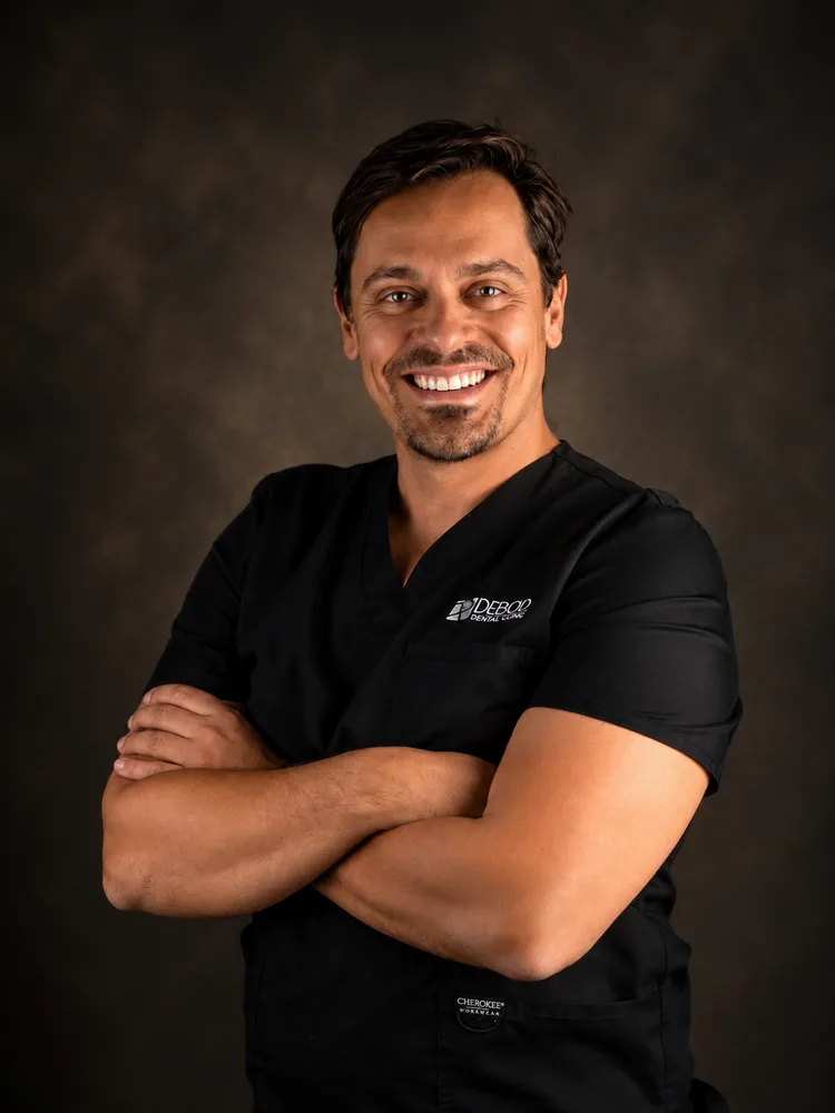
Author
Dr. César Rodríguez
Prosthodontics · Full Oral Rehabilitation
Dr. César Rodríguez is a distinguished prosthodontist with a Master's in Facial Prosthetics and Occlusion from the Complutense University of Madrid. Initially trained in Dentistry at the Central University of Venezuela, his credentials have…
Meet the team
Did this article raise questions?
Talk directly with our team of specialists. First consultation with no obligation.
Book a consultation